Dual Luciferase Assay Reagent for Gaussia luciferase - Red Firefly Luciferases
Please call for special pricing on bulk reagent purchase. Description:DLAR-1: Single Solution-based Dual Luciferase Assay Reagent: Save on costs and time in screening applications ) A panel of improved ultrasensitive secreted luciferase reporters have been developed in an effort to enable analysis of different promoter activities in the same group of transfected cells. This approach not only enables analysis of three or more pathways (responses) in the same group of cells but also enable one to study the response in real time without killing the cells since the three reporters are secreted. By choosing luciferases with different emission maxima we provide an additional advantage in that multiple luciferases can be assayed using a single assay reagent and spectrally resolving the two luciferase activities using appropriate filters. The single-solution based DLAR-1 dual assay reagent is based on the luciferase reporters enlisted below: Gaussia luciferase: Gaussia Luciferase is a luciferase from the marine copepod Gaussia princeps (1,2). This luciferase, which does not require ATP, catalyzes the oxidation of the substrate coelenterazine in a reaction that produces light, and has considerable advantages over other luminescent reporter genes such as secretablity and a much brighter signal intensity iaddition to excellent stability of the bioluminescent signal (approximately 10% decay in an hour. The luminescence measured from the supernatant of cultured cells transfected with a plasmid expressing GLuc is proportional to the amount of enzyme produced, which in turn, reflects the level of transcription. Alternatively a cell lysate can be used for the assay. Although most of the activity is secreted, measurements can be made from the cell lysates as well. A Red-emitting luciferase from the Italian firefly Luciola Italica. The emission wavelength of the red Luciola luciferases (617 nm). The robust signal of our Luciola luciferase mutants (1000 times higher signal intensity compared to native Luicola luciferase) makes them attractive for single solution –based multiplexed assays as signall strength is typically diminished in single solution-based multiplexed assays wherein different luciferse asctivities are spectrally resolved using appro priate filters. 

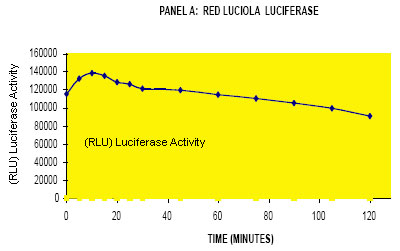
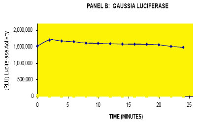
Figure 1: Stability of the bioluminescent signal of Gaussia Luciferase (Panel B) and Firefly luciferase (Panrl A) using the DLAR-1 reagent. This reagent is useful for HTS applications involving both Gaussia luciferase and the red-emitting Luciola luciferase. Note: Data presented is average of triplicate determinations measured on a Turner TD2020 luminometer. Figure 2: Emission spectra of Gaussia luciferase and Firefly luciferases in samples of transfected cells (lysates or supernatants). The emission spectra were recorded on a Fluorolog-3 spectrofluorometer (Horiba Scientific, Japan) using a liquid nitrogen cooled CCD. The luciferases were assayed by mixing 200 ul of the sample with the appropriate luciferase assay reagent to obtain spectral profiles. Emission max of Cypridina Luciferase is 463 nm; Red italica 617 nm. (Data courtesy of Justin Rosenberg, Dr Bruce Branchini´s lab, Connecticut College, USA) Advantages:
Assay Protocol for DLAR-1:Gaussia-Red Luciola luciferase dual assay reagent. Kit Contents:
Assay Protocol: Make sure all buffers are at room temperature prior to assay
Intracellular Gaussia luciferase activity Lyse cells using our lysis buffer (Catalog no 5X CLR-01). Follow cell lysis protocol supplied with the product. Assay as above using 5 ul to 10 ul of lysate NOTE: If you need to measure intracellular luciferase activity, lyse cells first using the cell-lysis buffer from Targeting Systems. (catalog no 5X CLR-01)
References:
Custom Reagents:Please check out our website www.targetingsystems.net for novel luciferase – based multiplexed assays which enable analysis of up to four promoter activities in the same group of transfected cells. |
