| Catalog no. |
Size |
Description |
Price |
| GAR-1 |
1000 assays |
Gaussia luciferase assay reagent |
$400.00 |
Please call for special pricing on bulk reagent purchase.
Description:
The GAR-1 Assay Kit is used for measurement of Gaussia luciferase activity in a standard (non-highthroughput format wherein samples can be measured quickly in a microplate reader or regular tube luminometer but long-term stability of the bioluminescent signal is not required. For applications such as high throughput screening we have developed assay reagents (GAR-2 and GAR-2B) which provide excellent stability of the bioluminescent signal due to incorporation of stabilizers. The GAR-2B reagent also provides a brighter signal intensity of the luciferase signal.
About Gaussia Luciferase:
Gaussia Luciferase (GLuc) is a luciferase from the marine copepod Gaussia princeps (1,2). This luciferase, which does not require ATP, catalyzes the oxidation of the substrate coelenterazine in a reaction that produces light, and has considerable advantages over other luminescent reporter genes such as secretablity and a much brighter signal intensity in addition to excellent stability of the bioluminescent signal (approximately 10% decay in an hour. The luminescence measured from the supernatant of cultured cells transfected with a plasmid expressing GLuc is proportional to the amount of enzyme produced, which in turn, reflects the level of transcription. Alternatively a cell lysate can be used for the assay. Although most of the activity is secreted, the high sensitivity of Gaussia luciferase allows measurements from the cell lysates as well.
Figure 1: Photo-oxidation of coelenterazine catalyzed by Gaussia luciferase
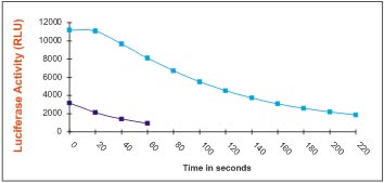
 Figure 2:
 Kinetics of of the GLuc bioluminescent signal using the GAR-1 reagent: Measurement of Gaussia luciferase activity in supernatants of cells (transfected with Gaussia luciferase expression vectors) using GAR-1 reagent from Targeting Systems (teal color line) and comparison with measurements obtained using the Renilla luciferase assay reagent form another commercial vendor (dark blue line).
Note: Data presented is average of triplicate determinations measured on a Turner TD2020 luminometer.

Advantages:
- Native Gaussia Luciferase (GLuc) possesses a natural secretory signal and upon expression is efficiently secreted into the cell medium. Cell-lysis not necessary for assaying the luciferase
- Gaussia Luciferase generates over 1000-fold higher bioluminescent signal intensity, than commonly used Firefly and Renilla Luciferases, making it an ideal transcriptional reporter (1).
- GLuc shows the highest reported activity of any characterized luciferases (1).
- The secreted protein is thermostable and has extremely high activity in light production allowing for very sensitive assays (1,2).
- The GLuc-containing samples (i.e. growth media or cell lysates after transfection) can be stored at -20°C for long-term storage.
 Assay Protocol:
 Make sure all buffers are at room temperature prior to assay
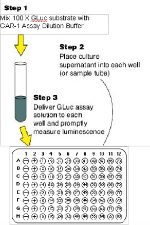
- Dilute the concentrated coelenterazine (GAR substrate), provided as a 100X formulation to 1X using the GAR assay dilution buffer. For example to prepare 5 ml of assay reagent dilute 50 ul of 100 X coelenterazine with 4.95 ml of GAR assay dilution buffer.
- Pipette 20µl of Gaussia Luciferase Sample into assay dish or luminometer tube
- Add 50 µl of Gaussia luciferase assay reagent (prepared as described in
step 1). Mix well
- 1.Assay in luminometer, integrate for 10 seconds (or as desired)
Intracellular Gaussia luciferase activity
NOTE: If you need to measure intracellular luciferase activity, lyse cells first using the cell-lysis buffer from Targeting Systems. (catalog no 5X CLR-01). Follow cell lysis protocol supplied with the product (see below). Assay as above using 5 ul to 10 ul of lysate
- Dilute the 5X CLR buffer 1:5 with water.
- Aspirate cell culture media and wash cells twice with serum free DMEM.
- Add enough of 1X cell lysis buffer to cover cells. Add enough lysis buffer to
cover cell.s (50 ul for 96-well, 300 ul for a 12-well, 800 ul for a 6-well dish
and 3 mll for a 10 cm dish
- Shake for 20 min at 400 rpm on an orbital shaker (room temperature).
- Mix 5-20 µl of luciferase containing sample or cell lysate with 50 µl of the
luciferase assay kit (GAR-1) and read immediately in the luminometer.
- All assay reagents should be close to room temperature at the time of
assay.
References:
- Tannous, B.A., Kim, D.E., Fernandez, J.L., Weissleder, R., and Breakefield, X.O. (2005) Mol. Ther, 11, 435-443.
- Wu, C., Suzuki-Ogoh, C. and Ohmiya, Y (2007) BioTechniques, 42, 290-292.
We can provide custom formulations to fit your HTS application.
Call our tech support team at 1-866-620-4018 or email us
info@targetingsystems.com or
targetingsystems@gmail.com
Please check out our website
www.targetingsystems.net for novel luciferase – based multiplexed assays which enable analysis of up to four promoter activities in the same group of transfected cells.


